Multiplexed Intra/extra-cellular imaging
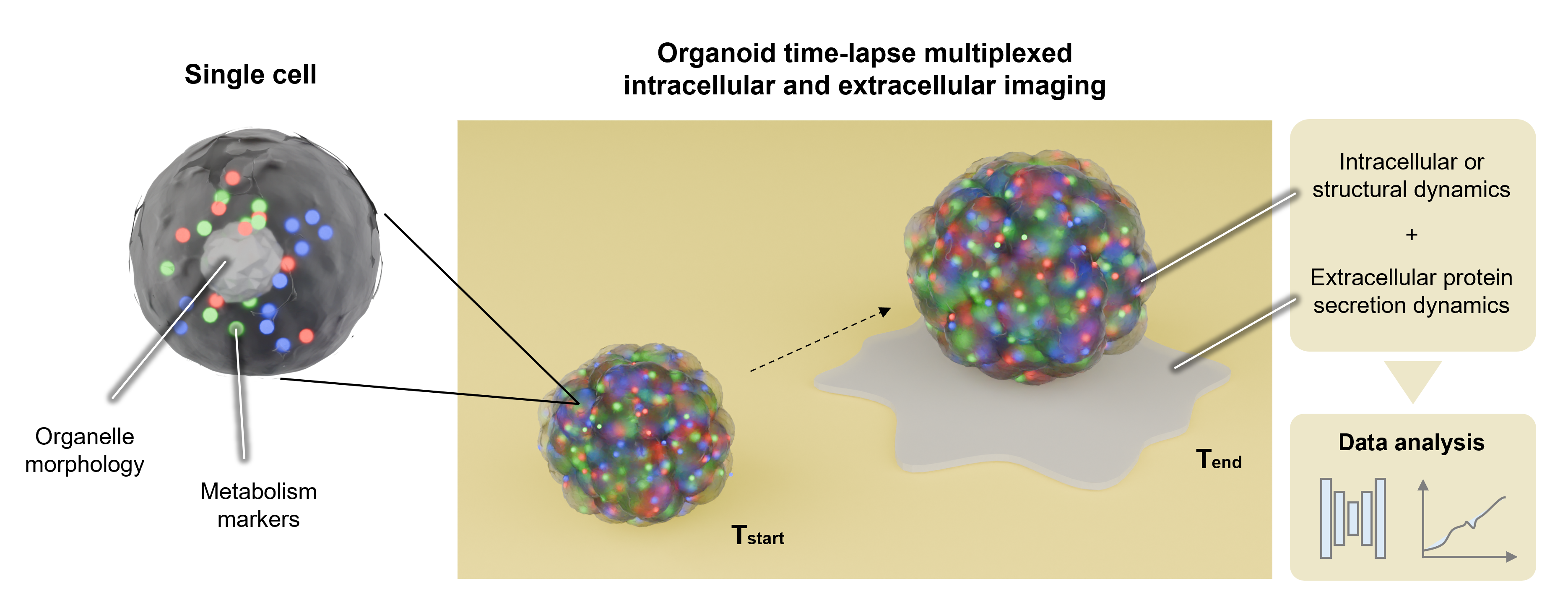
An optical biosensing project focusing on the development of a multi-model imaging system for simultaneously monitoring extracellular and intracellular dynamics in single cells and single organoids.
Optical imaging has been a powerful way to readout physiological and pathological information from biological samples. With the advancement of fluorescent microscopy and label-free imaging techniques, nowadays we can selectively visualize live-cell dynamics with fairly good spatiotemporal resolution. However, there is few methods that can simultaneously monitor intracellular and extracellular dynamics, leaving a knowledge gap in how biological systems coordinate these two distinct functional domains for cellular homeostasis and disease progression.
To enable new discoveries in this field, we developed a novel multi-model microscopy system combining labelled and label-free imaging for concommitant quantification of intracellular and extracellular dynamics with subcellular spatial resolution and minute-level temporal resolution. We demonstrated our imaging system using both single cells and organoids. We showcased a set of extracellular target and intracellular marker pairs, indicating the versatility and compatibility of the experimental workflow. To facilitate multi-dimensional bioimage analysis, we also established a deep-learning augmented image processing workflow and performed single-cell/organoid analysis in a high-throughput manner.
The newly-discovered phenotypic information has great potential in several translational applications, such as:
- Identifying heterogeneity of secretion dynamics and upstream intracellular regulators.
- Analyzing patient-derived samples as part of the personalized medicine measurements.
- Isolateing and sorting out single cell/organoid with selected secretion and morphological characteristics.

More information would be available after the manuscript get published. Stay tuned.